Loculated Pleural Effusion Cxr - Chest X-ray Shows Cardiomegaly With Infiltration And ... / Determining the cause of a pleural effusion is greatly facilitated by analysis of the pleural fluid.
Loculated Pleural Effusion Cxr - Chest X-ray Shows Cardiomegaly With Infiltration And ... / Determining the cause of a pleural effusion is greatly facilitated by analysis of the pleural fluid.. Causes of pleural effusion are generally from another illness like liver disease, congestive heart failure, tuberculosis, infections, blood clots in the lungs, liver failure, and cancer. Approximately 1 million people develop this abnormality each year in the united states. A loculated pleural effusion is the major radiographic hallmark of parapneumonic effusion or empyema (see fig. Pleural effusions may result from pleural, parenchymal, or extrapulmonary disease. A pleural surface permeability exudative effusion congestive heart.
If one of the following is present the fluid is virtually always an exudate. Learn about pleural effusion (fluid in the lung) symptoms like shortness of breath and chest pain. Pleural effusion occurs when too much fluid collects in the pleural space (the space between the two layers of the pleura). Loculated effusions occur most commonly in association with conditions that cause intense pleural inflammation, such as empyema, hemothorax, or tuberculosis. Pleural fluid ldh > two thirds of upper limit for serum ldh.
(A) Chest PA (before treatment): loculated right pleural ... from www.researchgate.net Large right effusion (red arrow) displacesthe heart to the left (yellow arrow). Bilateral pleural effusions withmeniscus signs. Pleural effusions can also loculate as result of adhesions. Loculated pleural effusion on cxr. It is commonly known as water on the lungs. It detects pleural effusions with higher sensitivity and specificity than cxr, and provides valuable information about the size and depth of the pleural effusion, the echogenicity of the fluid, the presence of septated or loculated fluid, pleural thickening and nodularity, and the presence of any. Tx if pt has chf. My pleural effusion healed without treatment.
Bhatia medical coaching institute, dbmci.
My pleural effusion healed without treatment. A pleural surface permeability exudative effusion congestive heart. The cardiac silhouette is also obscured. Differentiation of loculated effusions from solid masses. More than one half of these massive pleural effusions are caused by malignancy; Learn about different types of pleural effusions, including symptoms, causes, and the pleura is a thin membrane that lines the surface of your lungs and the inside of your chest wall. Pleural effusions can loculate as a result of adhesions. If none is present the fluid is virtually always a transudate. Bhatia medical coaching institute, dbmci. Approximately 1 million people develop this abnormality each year in the united states. Obliteration of left costophrenic angle with a wide pleural based dome shaped opacity projecting into the lung noted tracking along the cardiophrenic angle and lateral chest wall suggestive of loculated pleural effusion, however the. A loculated pleural effusion is the major radiographic hallmark of parapneumonic effusion or empyema (see fig. Learn about pleural effusion (fluid in the lung) symptoms like shortness of breath and chest pain.
Pleural fluid/serum ldh ratio >0.6. Large right effusion (red arrow) displacesthe heart to the left (yellow arrow). Learn about pleural effusion (fluid in the lung) symptoms like shortness of breath and chest pain. Dr bhatia discussing on pleural effusion in #lastminuterevisionpointdiscussionseries. Tx if pt has chf.
Massive effusion on left from www.lumen.luc.edu Pleural effusion symptoms include shortness of breath or trouble breathing, chest pain, cough, fever, or chills. It detects pleural effusions with higher sensitivity and specificity than cxr, and provides valuable information about the size and depth of the pleural effusion, the echogenicity of the fluid, the presence of septated or loculated fluid, pleural thickening and nodularity, and the presence of any. When you have a pleural effusion, fluid builds. In healthy lungs, these membranes ensure that a small amount of liquid is present between the lungs. Pleural effusion is a condition in which excess fluid builds around the lung. Determining the cause of a pleural effusion is greatly facilitated by analysis of the pleural fluid. Large right effusion (red arrow) displacesthe heart to the left (yellow arrow). Pleural effusion (imaging) introduction 1.
Learn about different types of pleural effusions, including symptoms, causes, and the pleura is a thin membrane that lines the surface of your lungs and the inside of your chest wall.
Determine if it can be tapped. Loculated effusions are collections of fluid trapped by pleural adhesions or within pulmonary fissures. A pleural surface permeability exudative effusion congestive heart. Case contributed by dr prashant mudgal. Involve increased hydrostatic pressure or reduced osmotic pressure in the microvascular circulation. Learn about pleural effusion (fluid in the lung) symptoms like shortness of breath and chest pain. Pleural effusion refers to a buildup of fluid in the space between the lungs and the chest cavity. Thoracentesis is a simple bedside procedure with imaging guidance that permits fluid to be rapidly sampled, visualized, examined microscopically, and quantified for chemical and cellular content. Detection of pleural effusion(s) and creation of initial differential diagnosis are a pleural effusion of 500 ml will obscure diaphragmatic contour on upright cxr; If none is present the fluid is virtually always a transudate. Dr bhatia discussing on pleural effusion in #lastminuterevisionpointdiscussionseries. Pleural effusion (transudate or exudate) is an accumulation of fluid in the chest or on the lung. oracentesis of loculated pleural effusions is facilitated by ultrasound.
Send aspirated fluid for cytology. Large right effusion (red arrow) displacesthe heart to the left (yellow arrow). Treatment depends on the cause. Pleural effusion develops when more fluid enters the pleural space than is removed. • congestive heart failure (40%):
Pleural Space Infections/Empyema - The Clinical Advisor from media.clinicaladvisor.com Computed tomography scan of the chest demonstrates loculated pleural effusion in the left major fissure (arrow) in a patient after coronary bypass. The pleura are thin membranes that line the lungs and the inside of the chest cavity and act to lubricate and facilitate breathing. Case contributed by dr prashant mudgal. There is a large left pleural effusion obscuring the lower half of the left hemi thorax. Bilateral pleural effusions withmeniscus signs. A pleural surface permeability) — exudative effusion. Pleural fluid/serum protein ratio >0.5. Pleural fluid ldh > two thirds of upper limit for serum ldh.
Detection of pleural effusion(s) and creation of initial differential diagnosis are a pleural effusion of 500 ml will obscure diaphragmatic contour on upright cxr;
Pleural effusion occurs when too much fluid collects in the pleural space (the space between the two layers of the pleura). Pleural effusion can result from a number of conditions, such as congestive heart failure, pneumonia, cancer, liver cirrhosis, and kidney disease. Send aspirated fluid for cytology. Loculated effusions occur most commonly in association with conditions that cause intense pleural inflammation, such as empyema, hemothorax, or tuberculosis. Detection of pleural effusion(s) and creation of initial differential diagnosis are a pleural effusion of 500 ml will obscure diaphragmatic contour on upright cxr; Pleural effusion is an accumulation of fluid in the pleural cavity between the lining of the lungs and the thoracic cavity (i.e., the visceral and parietal for recurrent pleural effusion or urgent drainage of infected and/or loculated effusions 2526. Approximately 1 million people develop this abnormality each year in the united states. A pleural surface permeability exudative effusion congestive heart. More than one half of these massive pleural effusions are caused by malignancy; Learn about pleural effusion including causes of pleural effusion. Thoracentesis is a simple bedside procedure with imaging guidance that permits fluid to be rapidly sampled, visualized, examined microscopically, and quantified for chemical and cellular content. Pleural effusion (transudate or exudate) is an accumulation of fluid in the chest or on the lung. A pleural effusion is accumulation of excessive fluid in the pleural space, the potential space that surrounds each lung.
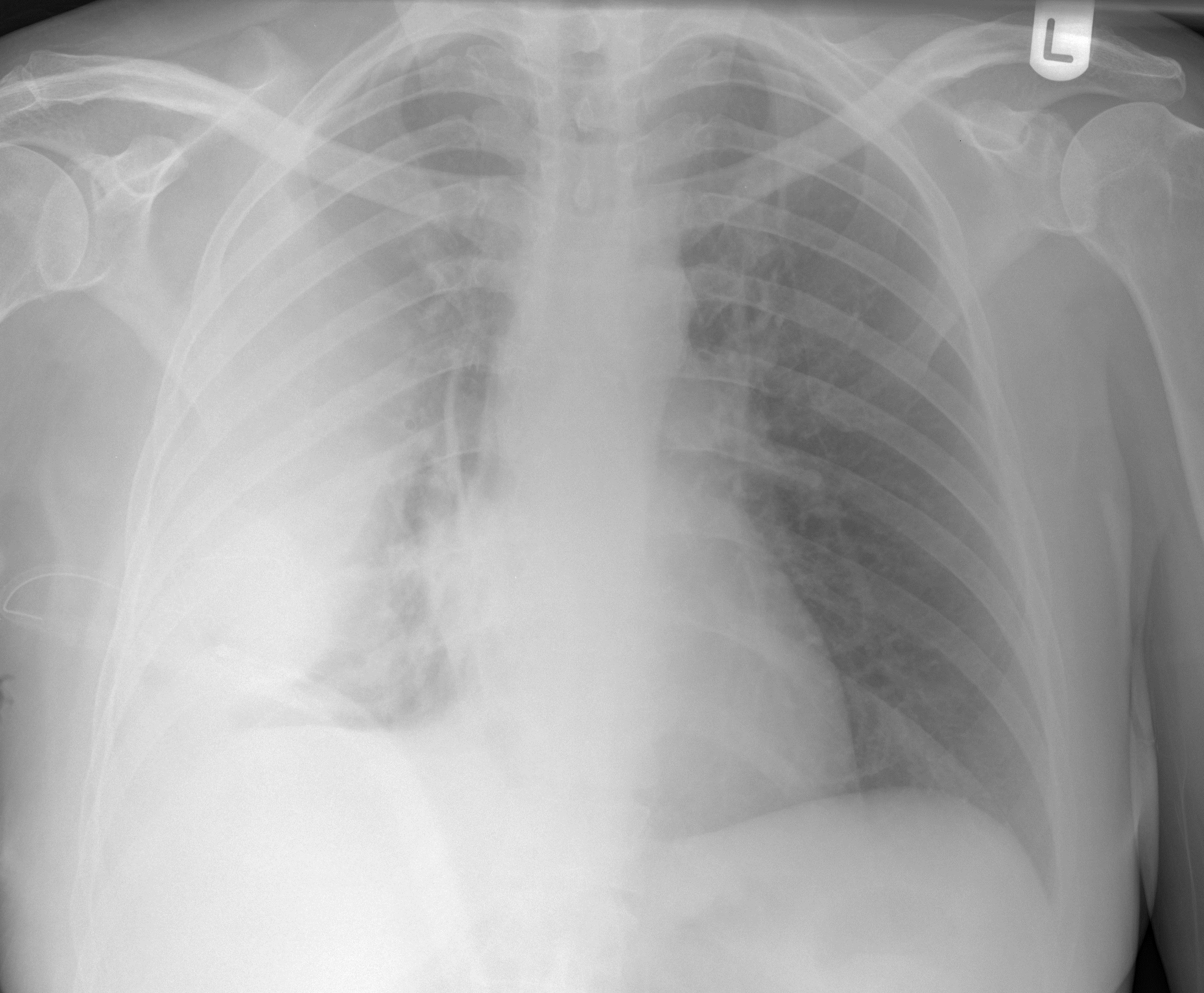
There is a large left pleural effusion obscuring the lower half of the left hemi thorax loculated pleural effusion. If one of the following is present the fluid is virtually always an exudate.


0 Komentar